Welcome to the first of a 2-part series on Left Ventricular Assist Devices (LVADs). Part 1 will provide an overview of LVADs, types of patients treated and the associated risks.
Congestive Heart Failure (CHF) is a leading cause of death worldwide. CHF is estimated to impact over 20 million people in the world, 2.2 million of which qualify for an LVAD. In the US alone, there are nearly 6 million people with CHF and 300,000 qualify for an LVAD.1
An LVAD is recommended when a patient reaches a stage of advanced heart failure when the heart is no longer able to pump enough blood to meet their body’s needs.2 The table below explains 3 crucial ways LVADs are utilized.
| 3 Uses of LVADs2 | |
| Bridge to Transplant | Implanted temporarily in a patient waiting for a heart transplant |
| Destination Therapy | Implanted as a permanent solution if a patient is not eligible for a heart transplant |
| Bridge to Recovery | Implanted for temporary heart failure when a heart may recover its strength after being given time to “rest” with the help of the device |
An LVAD is a surgically implanted mechanical pump that is attached to the heart. An LVAD works with the heart to help it pump more blood from the left ventricle to the aorta and, finally, throughout the body.
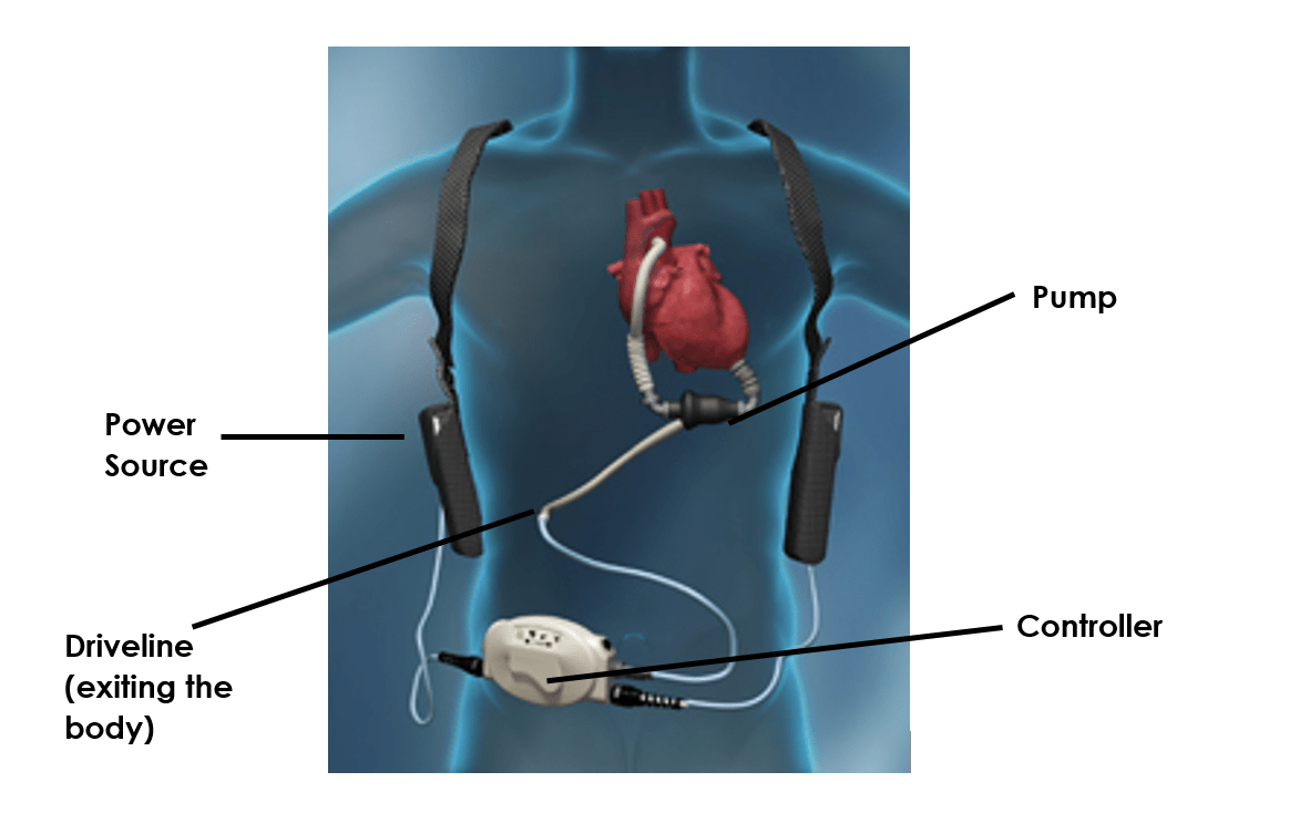
There are internal and external components of an LVAD which are highlighted in Figure 1.3

The pump provides blood flow, while the controller delivers power from the power sources (usually an external battery) to regulate the pump’s rate, sound alerts and display alarms if the system needs attention. The driveline exits the abdomen and attaches to the system controller, which regulates the LVAD’s function. This exit site requires meticulous, sterile dressing changes after the site heals and the patient returns home. The driveline must be securely immobilized to prevent irritation at the site to avoid infection and to prevent the wires from being damaged during regular activities.3
While LVADs have transformed treatment of advance heart failure, infection remains a significant risk.4 Consequently, the impact on clinicians, such as LVAD Coordinators and Infection Preventionists, is significant. Protection of the driveline that exits the patient’s body and connects the LVAD to the controller is critical.
Driveline infections are the most common type of LVAD-associated infection4, and these infections can be local, with pocket infection and driveline infection or they can be systemic, with involvement of the blood stream and/or endocarditis.5
In one study of 2006 patients receiving LVADs, percutaneous site infections (PSIs), occurred in approximately 19% of LVAD recipients by 1 year after implant and were associated with an increased risk of mortality.6
LVAD-associated infections likely begin with a disruption or trauma to the barrier between the skin and driveline. PSIs are commonly believed to involve the formation of a biofilm that make it difficult to eradicate bacteria.7
Several published studies have raised doubt about the durability of dressings (See Blog 13 for more details) The need to ensure the integrity of driveline dressings between scheduled changes has been established. The superior adhesiveness of Mastisol® Liquid Adhesive has been clinically demonstrated to improve the durability of dressings. Using Mastisol®:
- Reduces the likelihood of dressing or device migration or accidental removal
- Minimizes the risk of infection by creating a lasting occlusive dressing barrier
Trauma at the exit site of the driveline promotes the onset and maintenance of an inflammatory process and local infections. Avoiding excessive mobilization of the driveline will likely reduce the incidence of infections at the exit site and improve the quality-of-life of the patient.8 The use of a gentle, effective adhesive remover can minimize this friction/pulling at the skin during dressing changes.
Prevent skin damage from repeated application and removal of LVADs with Detachol® Adhesive Remover. Detachol® can play a significant role in assuring clinicians that they are doing everything possible to reduce the risks to patients and costs to healthcare providers that may result from the improper removal of tapes, dressings, and devices.
Using Detachol®:
- Reduces the risk of MARSI9
- Removes adhesive residue/bacteria10,11
- Provides a non-irritating, alcohol/acetone-free formulation
- Offers chlorhexidine gluconate (CHG) compatibility12
Dressing integrity is crucially important once an LVAD patient returns home. Because patients and/or their caregivers are responsible for maintenance of their LVAD once at home, proper training of dressing maintenance as well as the risks of non-adherent dressings is essential. Follow your hospital or clinic’s protocol for specific directions and recommendations for exit site care.
For more information about Mastisol®, please contact your sales consultant or Eloquest Healthcare®, Inc., call 1-877-433-7626 or visit www.eloquesthealthcare.com.
Minimizing infection risk is an essential part of optimizing “The Triple Aim” of the Affordable Care Act. Eloquest Healthcare is committed to providing solutions that can help you reduce the risk of conditions like a central line-associated bloodstream infection (CLABSI) and post-operative wound contamination.
Return for more tips and specifics in part 2 of the series, “LVADs: Minimizing Driveline Infections at Home.”
References: 1. HRF. https://healthresearchfunding.org/25-fascinating-lvad-statistics/. Accessed 01/11/18. 2. MyLVAD. https://www.mylvad.com/content/what-lvad-how-does-it-work. Accessed 01/11/2018.
3. Bond AE, Bolton B, Nelson K. Nursing education and implications for left ventricular assist device destination therapy. Prog Cardiovasc Nurs. 2004;19(3):95-101. 4. Leuck AM. Left ventricular assist device driveline infections: recent advances and future goals. J Thorac Dis. 2015;7(12):2151-2157. 5. Hernandez GA, Breton JD, Chaparro SV. Driveline infection in ventricular assist devices and its implication in the present era of destination therapy. Op J Card Surg. 2017;9:1-6. 6. Goldstein DJ, Naftel D, Holman W, Bellumkonda L, Pamboukian SV, Pagani FD, Kirklin J. Continuous-flow devices and percutaneous site infections: clinical outcomes. J Heart Lung Transplant. 2012; 31(11):1151-7. 7. Trachtenberg BH, Cordero-Reyes A, Elias B, Loebe M. A review of infections in patients with left ventricular assist devices: prevention, diagnosis and management. Methodist Debakey Cardiovasc J. 2015;11(1): 28–32. 8. Baronetto A, Centofanti P, Attisani M, Ricci R, Mussa B, Devotini R, Simonato E, Rinaldi M. A simple device to secure ventricular assist device driveline and prevent exit-site infection. Interact Cardiovasc Thorac Surg. 2014;18(4):415-7. 9. McNichol L, Lund C, Rosen T, et al. Medical adhesives and patient safety: state of the science. Consensus statements for the assessment, prevention, and treatment of adhesive-related skin injuries. J Wound Ostomy Continence Nurs. 2013;40:365-80. 10. Berkowitz DM, Lee W-S, Pazin GJ, et al. Adhesive tape: potential source of nosocomial bacteria. Appl Microbiol. 1974;28:651-54. 11. Redelmeier DA, Livesley NJ. Adhesive tape and intravascular-catheter-associated infections. J Gen Intern Med. 1999;14:373-5. 12. Ryder M, Duley C. Evaluation of compatibility of a gum mastic liquid adhesive and liquid adhesive remover with an alcoholic chlorhexidine gluconate skin preparation. J Infus Nurs. 2017;40(4):245-52.


